Biodiversity Research and Teaching Collections’ oVert project on display at Texas A&M

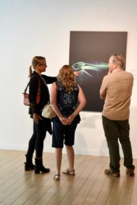
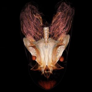
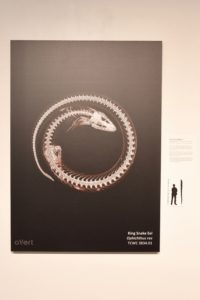
COLLEGE STATION — The Biodiversity Research and Teaching Collection, or BTRC, maintained by the Texas A&M Department of Wildlife and Fisheries Sciences has launched a gallery of images created using CT scans from their collection of specimens, including a bigeye thresher shark, an alligator snapping turtle, a clingfish and three other images.
The gallery, on display from June 12 to Sept. 21, is hosted by the Reynolds Gallery at the Memorial Student Center at Texas A&M University, 275 Joe Routt Blvd., College Station.
The gallery exhibit, which is free and open to the public, is part of the “Open Vertebrate Exploration in 3D” project, or oVert. Texas A&M is one of 16 institutions involved in the multimillion-dollar project backed by the National Science Foundation, or NSF.
Using specialized scanners designed for human and veterinary medical uses, the BRTC along with the Texas A&M Institute for Preclinical Studies and the Texas A&M College of Veterinary Medicine are working together to scan some of the largest specimens in the project.
Six examples of these high-quality digital scans are on display in the Reynolds Gallery, organized by Heather Prestridge, curator of the BRTC, and Mary Compton, curator of the Reynolds Gallery.

“Some of these animals are only preserved in a few collections around the world, which makes them difficult and sometimes impossible to study in detail,” Prestridge said. “With digital scans that can be shared electronically, scientists have access to thousands of unique animal species at their fingertips.”
“Each scan comes with its own story,” Prestridge said. “There is a skull of a fish that’s inside this eel that he ate, but we would have never known that if we didn’t CT scan it. So now we have colleagues at the University of Florida working on segmenting this piece out so we can identify the fish.”

Each image is accompanied by a description and a scaled image to give viewers a visual depiction of the size of the specimen. The specimens range from a few inches to a few feet in length.
“Nothing that we do is standard,” Prestridge said. “Each is a different size. Each is a different shape. This makes our job challenging and forces innovation.”
Each image on display is brightly colored in different gradients that are strictly related to the density of the material and is used purely as an artistic application, she explained.
The goal of the project is to scan more than 20,000 unique animal species by 2021. The best specimens are identified, and then their digital images are uploaded to a free online database called MorphoSource, she explained.
“Half of our download requests are for ‘non-research’ use,” Prestridge said. “This helps collections like ours to engage non-traditional user groups, including K-12 education, which can use our data to 3D print specimens and artists who work in digital media.”


