Texas A&M researchers peer into the microcosmos with high-resolution microscope
Powerful cryogenic-electron microscope reveals interactions at the molecular scale
Here’s a movie trivia question for you: How did the scientists in “Jurassic Park” retrieve the DNA they used to clone the dinosaurs?
If you grew up with those movies, you might remember the zany description of how the cloning was made possible: “A hundred million years ago, there were mosquitoes, just like today. And, just like today, they fed on the blood of animals, even dinosaurs. Sometimes, after biting a dinosaur, the mosquito would land on the branch of a tree and get stuck in the sap…”
By extracting the blood from a mosquito trapped in amber, the scientists obtained the genetic blueprint to clone dinosaurs. And, while that idea is itself trapped within the realm of science fiction, Junjie Zhang, Ph.D., associate professor in the Department of Biochemistry and Biophysics, said it’s fairly similar to freezing a sample for observation with a cryogenic-electron microscope, or cryo-EM.
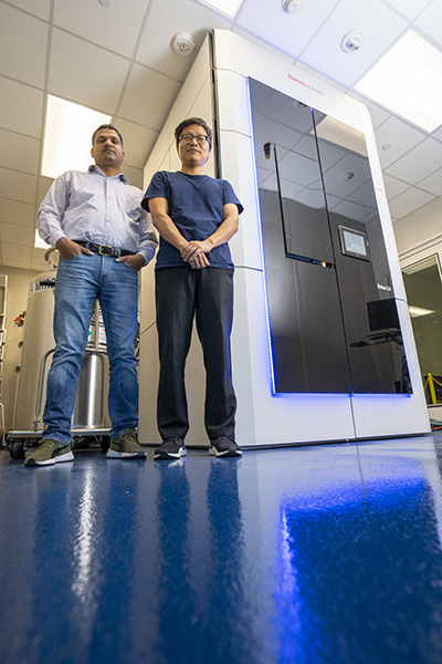
Within the Texas A&M College of Agriculture and Life Sciences, researchers are resolving some of the tiniest biological and chemical structures and viewing detailed molecular interactions by cryogenically freezing samples in place, capturing them like a mosquito in amber. Once the sample is frozen, the scientists can use the high-resolution electron microscope to observe what’s happening at the molecular scale.
“Instead of using amber, we use a special kind of ice called vitreous ice that doesn’t disrupt the proteins or nucleic acids very much,” Zhang said. “Similar to how the amber protects and preserves the mosquito, the ice preserves the molecules or cells so that we can image the structures and interactions.”
Unlike the “Jurassic Park” movies, these scientists won’t be bringing any huge man-eating animals back from extinction. But they are answering some questions that hold huge biological implications for the world on everything from infectious disease and structural biology to cancer.
Who’s using the cryo-EM at Texas A&M
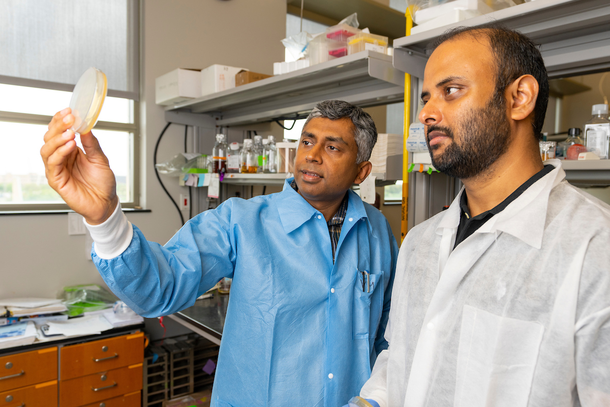
Since the cryo-EM’s initial opening in November 2022, it’s already aided in several high-impact research projects. For instance, it was only with the use of the cryo-EM that Zhang and Lanying Zeng, Ph.D., also a professor in the Department of Biochemistry and Biophysics, were able to detail the mechanism of how a virus infects a bacterial cell by attaching to an appendage on the cell surface.
“We can use the cryo-EM to literally see interactions between a cell and bacteriophage,” Zhang said. “Other imaging techniques wouldn’t give a high enough resolution. They wouldn’t tell you which part of the virus is interacting with which part of the bacteria or show precise physical interactions.”
However, using the cryo-EM, Zhang said they can see the interface between a protein on a bacteriophage and another on a bacterium.
“And with that, it allows us to understand exactly how the phage recognizes its host and eventually destroys it,” he said.

Other researchers on campus are also advancing their own fields with the help of the cryo-EM. Along with scientists within Texas A&M AgriLife Research and the College of Agriculture and Life Sciences, Zhang said researchers within Texas A&M’s College of Arts and Sciences, College of Engineering, School of Medicine and School of Pharmacy have used the microscope for various projects.
Zhang said having local access to the wide-ranging capabilities of the cryo-EM has made it a powerful tool for Texas A&M researchers — and it’s also proven to be a focal point for scientific collaboration across the state of Texas.
Beyond Bryan-College Station, Gaya Yadav, Ph.D., technical director of the cryo-EM, oversees image collection requests from Baylor College of Medicine, Rice University, Texas Tech University, the University of Texas at Arlington and the University of Texas MD Anderson Cancer Center in Houston.
Researchers can submit their samples for imaging by the cryo-EM staff, or trained researchers can schedule a visit to campus and use the facility themselves with oversight from Yadav.
“This is the first microscope of this kind within The Texas A&M University System, and we’re very fortunate to have it available for impactful research on campus,” said Josh Wand, Ph.D., University Distinguished Professor and head of the Department of Biochemistry and Biophysics. “The operation of our microscope is already at almost 100% capacity, and we’ve received excellent feedback from our external users, who have all been very satisfied with our staff’s service and expertise.”
What makes the cryo-EM so powerful
When asked to picture a microscope, the image that comes to mind for most people would probably be a light microscope. These are the standard microscopes sitting on lab benches all over the world.
Those light microscopes are invaluable for everyday use, but, unfortunately, the limit of their resolution leaves much unseen.
Zhang said even the most powerful versions of light microscopes run into a limit because of the wavelength of light. A light microscope’s resolution can distinguish between as few as 0.2 micrometers. For perspective, the breadth of a human hair is around 100 micrometers. So, if a scientist wanted to examine a protein structure, which might be a thousand times smaller than a micrometer, light microscopy is out of the question.
An electron microscope, on the other hand, can resolve sample images at less than one billionth the size of a meter. In the past few years, the technology has advanced enough that individual atoms can be localized in a protein structure.
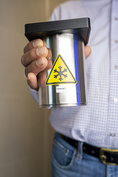
To image a sample with the cryo-EM, scientists rapidly freeze a small volume at a temperature around minus 300 degrees Fahrenheit. The rapid freezing changes the water in the sample to vitreous ice, which is water that freezes so quickly it’s unable to form a normal crystalline ice structure. Zhang said this step preserves the sample’s natural physiological conditions, which many other techniques would disrupt by staining, dehydrating or crystallizing.
A machine then shoots a beam of electrons at the sample to create thousands of images showing different perspectives of the target molecule, which can then be used to create a structural model.
This is an example of single-particle cryogenic-electron microscopy. Zhang said the facility at Texas A&M is also capable of cryo-electron tomography, which allows for imaging of intact cellular structures in their natural state, offering insights into the spatial organization and architecture of entire cells.
Continued growth and plans for expansion
In the spirit of boundless science, Zhang said the team has already upgraded since the cryo-EM’s debut to include a cryo-confocal fluorescent microscope, which combines the benefits of cryogenic freezing with the molecular-tagging capabilities of light microscopy. The team is also seeking external funding to advance the facility’s cryo-electron tomography imaging power.
“At Texas A&M AgriLife, collaboration and pushing the boundaries of science are fundamental to our mission,” said Jeffrey W. Savell, Ph.D., vice chancellor and dean for Agriculture and Life Sciences. “With convenient access to the state-of-the-art cryo-EM, our researchers uncover new insights into the microscopic world and direct macroscopic advances across the life sciences and human health.”
Here’s hoping those advances steer clear of pre-historic carnivores.